New Research on the Sensitivity of Elephants’ Trunks

An elephant’s trunk is even more sensitive than initially thought
We know that elephants use their trunks for multiple purposes – drinking, feeding themselves, feeling objects – but a new research paper published last month indicates that the neurons controlling the sense of touch in an elephant’s trunk number 400,000, far more than had been expected. This means that elephants are likely to be even more tactile and rely even more on their sense of touch than had originally been thought.
This preliminary study was carried out by a team of scientists (see Current Biology, doi.org/hdb2) who dissected the heads of elephants that lived in zoos and died naturally or as a result of disease. They discovered that the elephant trigeminal ganglion (the cranial nerve responsible for transmitting sensory information to the brain) is one of the largest known primary sensory structures. It also contains very large neurons. These factors, together with the high number of neurons in the infraorbital nerve, indicate a high degree of tactile specialisation in elephants and it is likely that they rely even more heavily on their sensory nerve than previously thought. Given that their infraorbital nerve (a branch of the trigeminal ganglia) is three times thicker than their optic nerve, they may even favour feel over sight when it comes to assessing what’s in front of them.
The authors noted that behaviour studies suggest a high degree of tactile sensitivity of the elephant trunk, and that elephants constantly touch the environment with their trunk. They comment that whereas such behaviour may seem manipulative to the human eye, it might serve as an important sensory function. The fact that the infraorbital nerve is thicker than the spinal cord leads to the suggestion that elephants are very tactile.
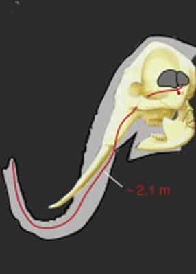
The first photograph below is a diagram (taken from the study) of an elephant head with the brain in dark grey and a schematic of a trunk sensory neuron (in red). The neuron’s cell body (red circular dot) is situated below the brain in the trigeminal ganglion.
[Click on photos below to enlarge]

Keep up to date. Sign-up to our newsletter now to be the first in the know, and get our latest news direct to your inbox.
